Finasteride may increase risk of Alzheimer's: "Inhibition of 5 α reductase impairs cognitive performance, alters dendritic morphology and increases tau phosphorylation in the hippocampus of male 3xTg-AD mice"

Highlights

• Inhibition of 5α reductase with finasteride impaired object recognition memory in 6 month- old male 3xTgAD (Alzheimer) mice.

• 5α reductase inhibition resulted in a two-fold increase in site-specific Tau phosphorylation in the hippocampus.

• CA3 apical dendritic branching and spine density also decreased in finasteride-treated 3xTgAD males.

Abstract

Recent work has suggested that 5α-reduced metabolites of testosterone may contribute to the neuroprotection conferred by their parent androgen, as well as to sex differences in the incidence and progression of Alzheimer’s disease (AD). This study investigated the effects of inhibiting 5α-reductase on object recognition memory (ORM), hippocampal dendritic morphology and proteins involved in AD pathology, in male 3xTg-AD mice. Male 6-month old wild-type or 3xTg-AD mice received daily injections of finasteride (50 mg/kg i.p.) or vehicle (18% β-cyclodextrin, 1% v/b.w.) for 20 days. Female wild-type and 3xTg-AD mice received only the vehicle. Finasteride treatment differentially impaired ORM in males after short-term (3xTg-AD only) or long-term (3xTg-AD and wild-type) retention delays. Dendritic spine density and dendritic branching of pyramidal neurons in the CA3 hippocampal subfield were significantly lower in 3xTg-AD females than in males. Finasteride reduced CA3 dendritic branching and spine density in 3xTg-AD males, to within the range observed in vehicle-treated females. In the CA1 hippocampal subfield, dendritic branching and spine density were reduced in both male and female 3xTg-AD mice, compared to wild type controls. Hippocampal amyloid β levels were substantially higher in 3xTg–AD females compared to both vehicle and finasteride-treated 3xTg–AD males. Site-specific Tau phosphorylation was higher in 3xTg-AD mice compared to sex-matched wild-type controls, increasing slightly after finasteride treatment. These results suggest that 5α-reduced neurosteroids may play a role in testosterone-mediated neuroprotection and may contribute to sex differences in the development and severity of AD.

9 Likes

Beauty.

2 Likes

Apologies can you help me understand?

This happens because some people have lowered testosterone?

Also, article says it progresses Alzheimer’s more, but not start it in non Alzheimer’s male rats?

I posted the paper on Reddit - Tressless. The level of denial is unbelievable.

2 Likes

you have a higher chance of convincing a brick wall not to use fin then those guys on hair loss forums

2 Likes

That’s what I suspected :confused:

Actually, I decided to get my brain analysed in case I die from something. I am sure they will find severe damage in the Hippocampus and some other areas of the brain.
It reminds me of the CTE (chronic traumatic encephalopathy) in football. Just a few years ago the NFL denied heavily that football leads to CTE which is an Alzheimer’s like disease. It needed the autopsy the brains of 100 dead football players to change this fact.
The same is happening with Merck (like NFL) und PFS (like CTE). Therefore in case something happens to me, I want some scientists to have a close look at my organs and how PFS could have affected them.

@lakehouse not testosterone, but testosterone cannot be converted well into molecules that are made from testosterone further downstream.

1 Like

so my DHT levels are normal, as is my free testosterone range.

is this enough information to see if im at risk for whats seen in the research? or is it a different mechanism that affects us all irregardless of hormone levels?

thanks for the info

The problem is no-one cares. They can look into your organs and tissues while you are still alive and they can find a lot of things but nobody is doing that. Nobody has tested your DHT or 5ar2 expression in prepuce, for example, or neurosteroids in spine fluid.

1 Like

Yes, they may not care. But I and my family do care. If paid for it privately l, any kind of test can be done.

Sure. But that won’t change the scientific consensus because it won’t be a “study”. An anecdotal report can be written and published though, which is still something.

There is no point to test or analyze the brain though. Science knows nothing about the brain so data cannot be interpreted.

Here are some things that could be tested, in my opinion:

I very much disagree that we know nothing about the brain and that it would yield valuable data.
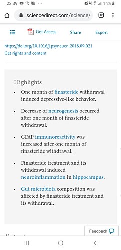
Melcangi described brain changes in rates after finasteride treatment and withdrawl. This are the results:

Showing similar results in real patients would be an important step.

I also expect microgliosis, increased beta amyloid plaques and tau as a sign of a neurogenerative process.

The fact that PFS patients (maybe just a subgroup) have altered hormones in spine fluid point to a pathological process in the brain which fits to the neurological symptoms in many.

I personally think that there are at least 2 subgroups of PFS patients

a) the one who describe changes in their genitals
b) the ones who may have more neurological symptoms

Probably in group b DHT in genitals is fine and no silencing in the 5argene could be found, but in the spine fluid DHT may be low and 5ar silenced
Probably in group a you would see the opposite.

This is just speculation, but there is a lot of reason think that 5ar is not silenced in every tissue in the body. Especially brain and the rest of the body are separated by the blood brain barrier. So you will have different results if someone is affected peripherally or primarily in the nervous system.

1 Like

Really unbelievable? On this forum, where of all places people should acknowledge different outcomes, a small percentage of posters still endlessly deny the clear symptoms/responses of others due to survivorship or confirmation biases. In my opinion this is less unbelievable and more unavoidable:

Re the study: More and more evidence is emerging regarding the important role of AR-mediated signaling in the hippocampal CA1 region with relevance to AD. You may find this interesting:

3 Likes

This is pretty fucking scary.